



UNITY® M Ophthalmic Surgical Microscope
Designed to balance the demands for enhanced visualization with exceptional precision,* control, and adaptivity.1-4
UNITY® M Ophthalmic Surgical Microscope
Designed to balance the demands for enhanced visualization with exceptional precision,* control,
and adaptivity.1-4
The Power of Precise Control1-3
Experience consistent visualization that supports controlled, confident surgery across cataract, cornea, retina, and MIGS procedures.1-3

Expansive Red Reflex
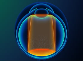
Expansive red reflex geometry with an area over 2000 mm2 minimizes the need to chase the eye,
maintaining continuous visualization during surgery.1

Expanded Depth of Field
Delivers a depth of field of approximately 3,200 microns at 7x magnification.† Average human anterior chamber depth is approximately 3 mm.2,5

Longer Maximum Illumination Exposure Time
Provides 4.6x longer maximum illumination exposure time vs. focused beam illumination.6,7‡

Advanced Mode for Posterior (AMP)
Delivers up to 42% more light to the surgeon without increasing illumination in the patient’s eye.8,9
Alcon’s UNITY® M is an ophthalmic surgical microscope built exclusively for ophthalmology.4


Precision

Experience a stable red reflex and a deep depth of field
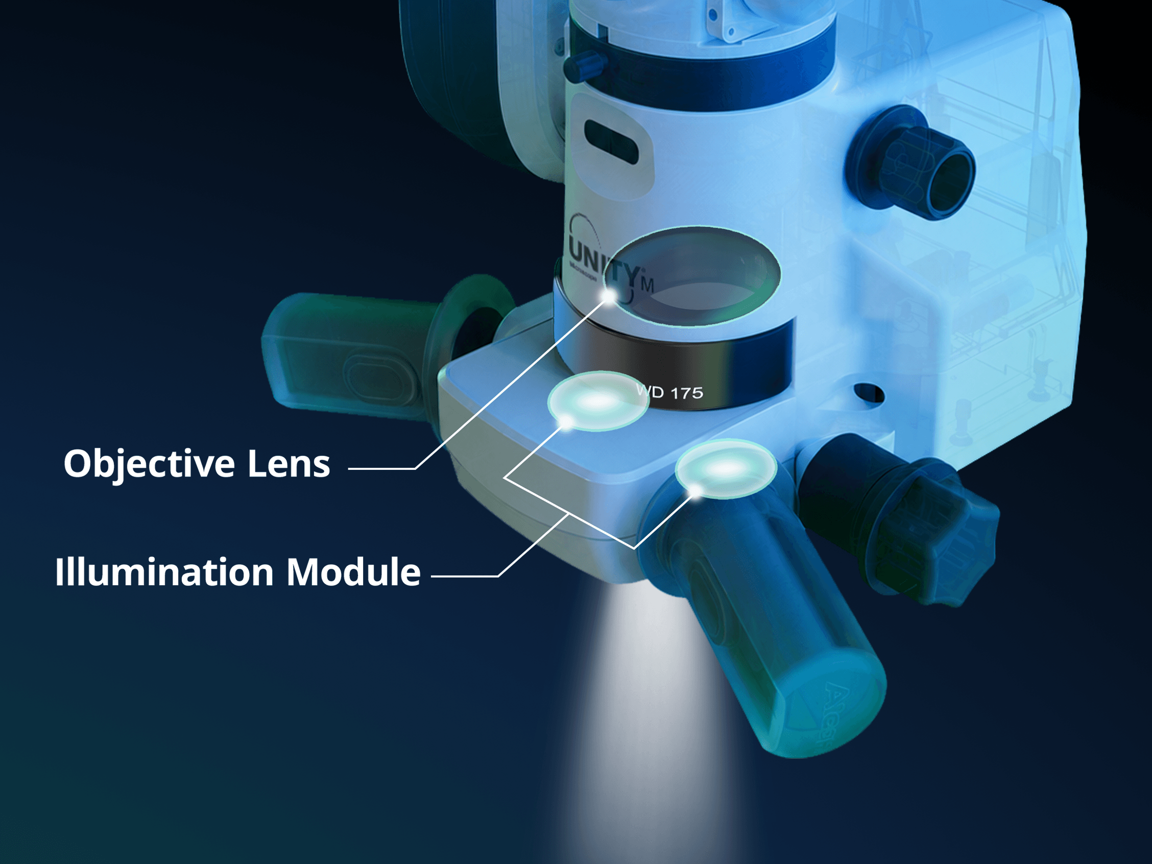
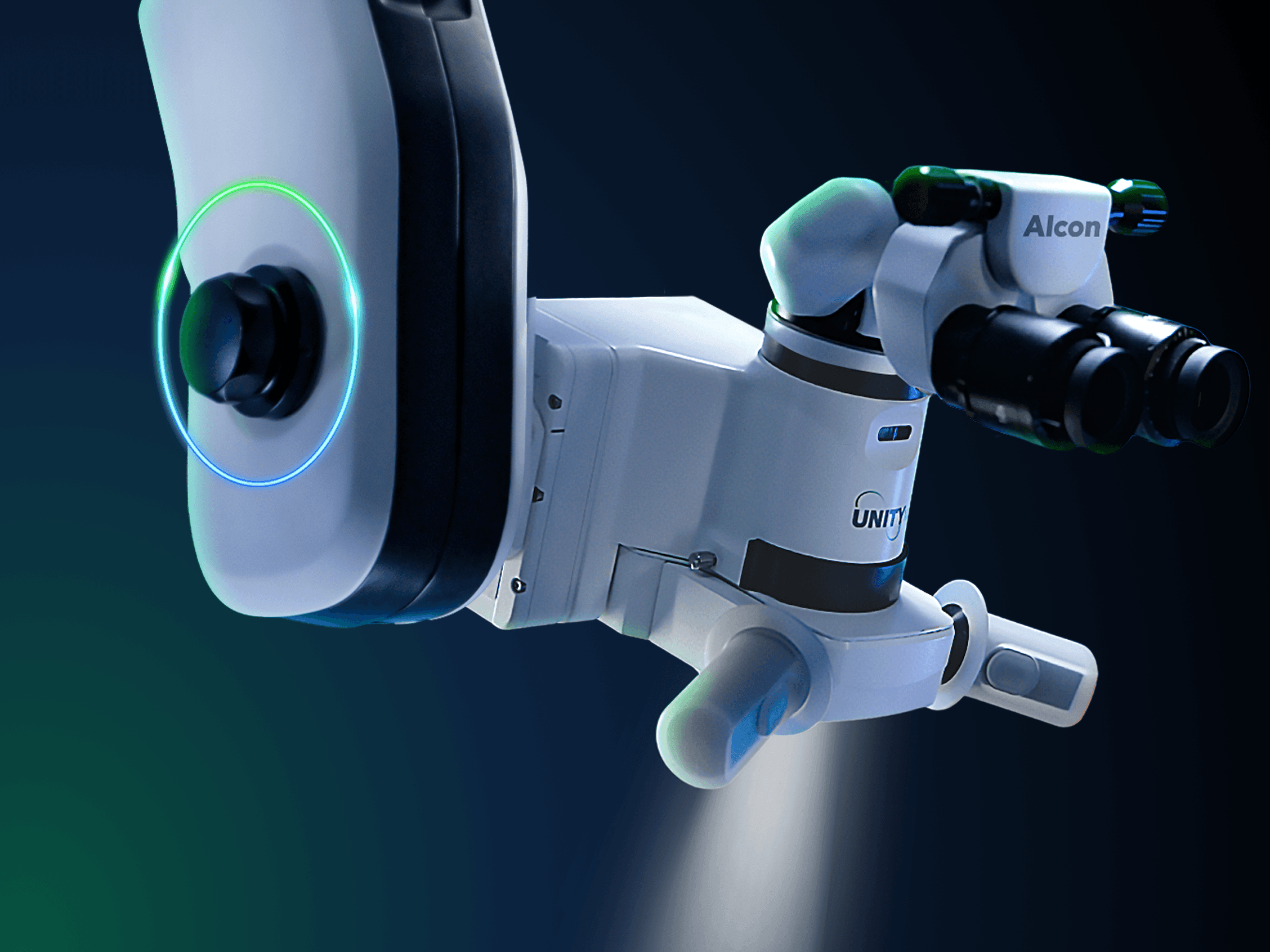
UNITY® M positions the objective lens above the illumination module which produces nearly collimated illumination and increases the focal length across working distances, helping deliver a stable red reflex and enhanced depth of field.1
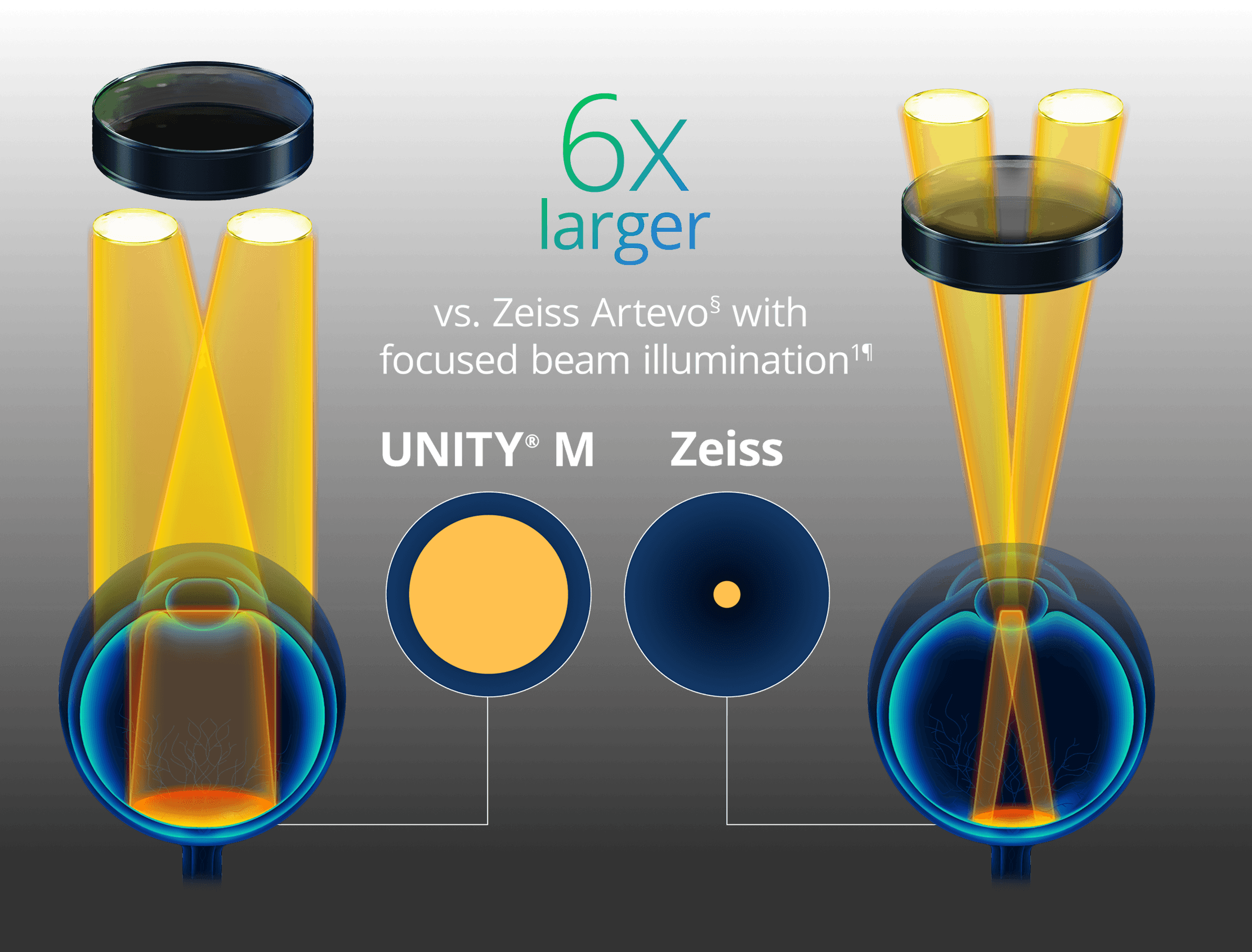
A red reflex that stays locked in1,10,11
Enabled by Superior Lens Position, nearly collimated
illumination delivers a bright, stable red reflex zone that
is less susceptible to pupil size, microscope position, or patient eye movement.1,11-13‖
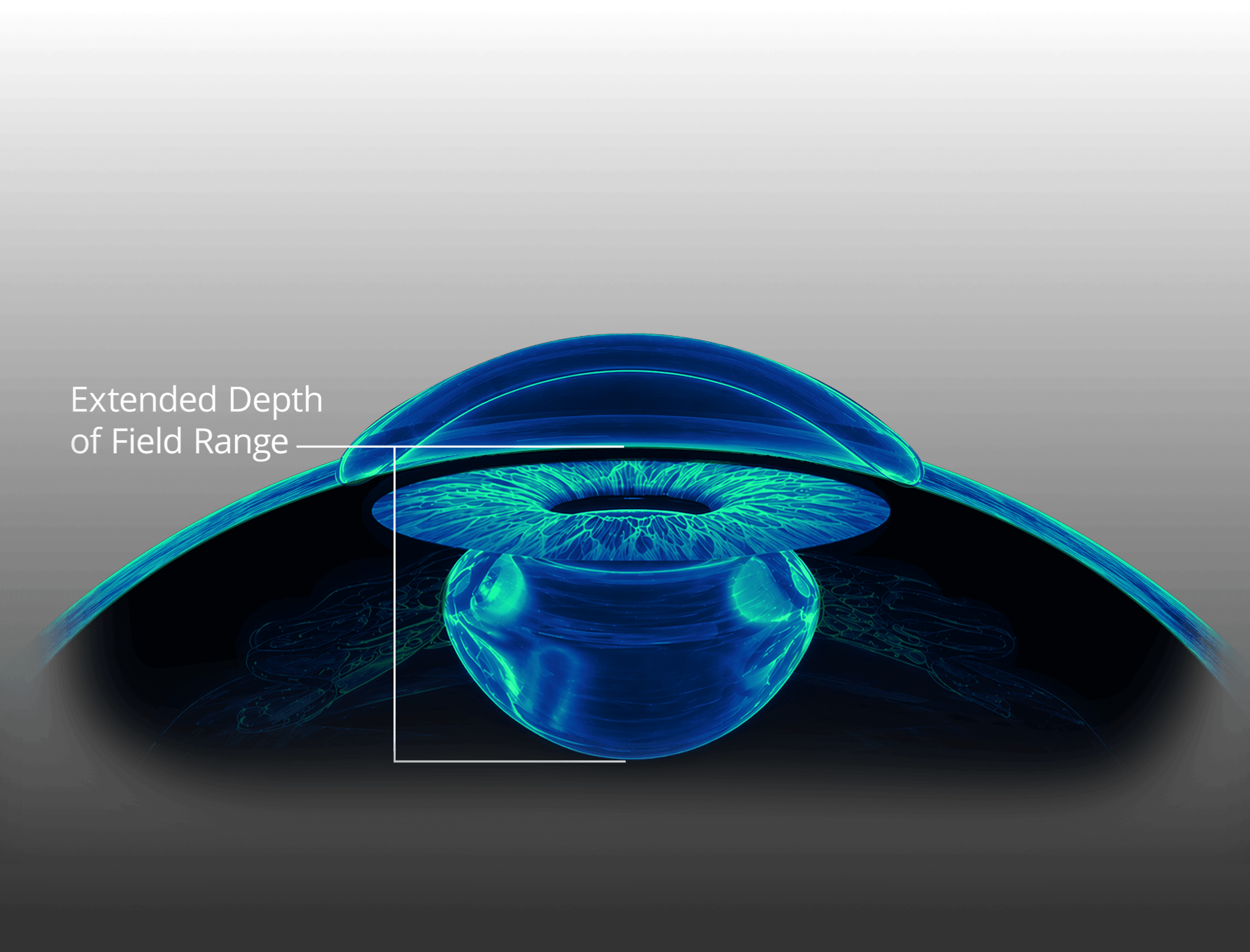
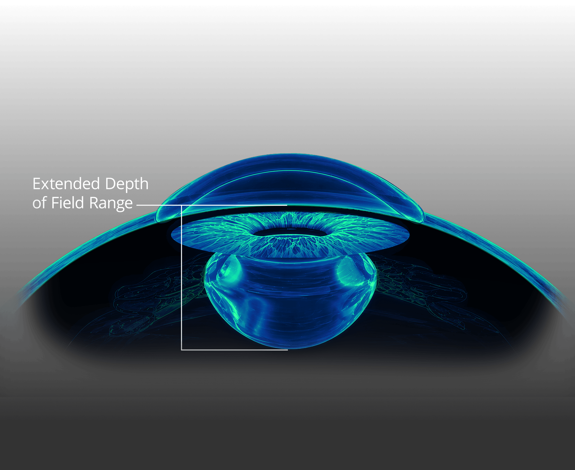
A depth of field that covers the chamber
UNITY® M delivers a depth of field of approximately 3,200 microns at 7x magnification†, supporting uninterrupted visualization of structures as the eye shifts or the chamber changes. Average human anterior chamber depth is approximately 3 mm.2,5

Control

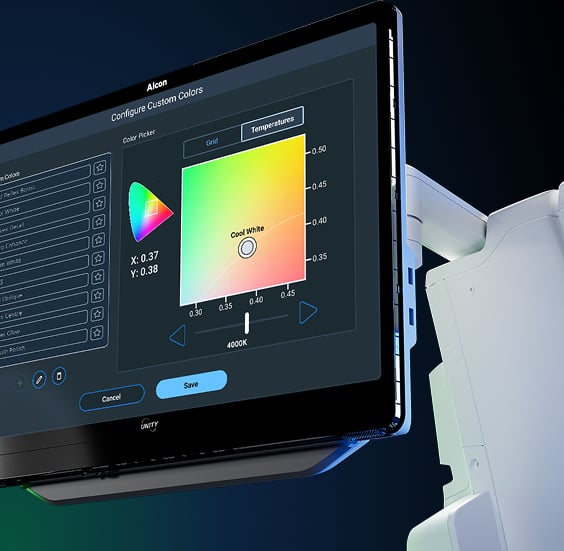
First and Only Microscope with Color Tunable Illumination7,14,15**
Control coaxial and oblique light independently—across Kelvin temperature and, for the first time, color spectrum.7,14,15
UNITY® M’s first and only Color Tunable LED Illumination delivers improved contrast and tissue detail, helping surgeons distinguish critical structures across ophthalmic procedures.2,7,14-16**†† Up to 16 different preset colors are available to choose from, with the flexibility to further customize illumination to your preference.
Longer Maximum Exposure Times

UNITY® M provides high-definition visualization at low light settings, reducing patient exposure to bright light during surgery. Longer safe exposure time helps minimize the risk of phototoxicity.6,7,10,17‡

UNITY® M has 4.6x longer maximum illumination exposure time vs. focused beam illumination.6,7‡
Light Sentry Mode
Minimize the risk of photochemical retinal damage during surgery.4
When Light Sentry mode is enabled, the maximum permitted illumination level is limited to 30% of the 100% output available.4
Extends maximum illumination exposure time to >100 minutes.4‡‡

Advanced Mode for Posterior
Deliver up to 42% more light to the surgeon without increasing illumination in the patient's eye.8,9
AMP removes the internal beam splitter from the optical path, enabling full transmission of light and supporting clear posterior visualization.8

Electronic Tilt for Controlled View
Adjust the optical head to a preset
or customized angle.4
Electronic tilt supports ergonomic comfort and ease of use during MIGS and other ophthalmic procedures.4
Built for a Wide Range of Ophthalmic Procedures
| Procedure | UNITY® M Visualization |
|---|---|
Cataract | Designed for modern cataract surgery with high resolution for high-precision steps (including capsule polishing).2 |
Cornea | Consistent clarity from capsule to cornea.2 |
Retina | Tissue structures and tool edges stay clearly defined.2 |
MIGS | The angle structures and instrument tip stay simultaneously crisp.2 |

Optimization
Modular by Design.
Built Around You.4
Designed to support practice-specific customization and future scalability.4
UNITY® M’s modular platform can be configured to surgeon preference and procedure type without the need to replace the entire system.4 A connectivity module supports current and future OR connectivity.
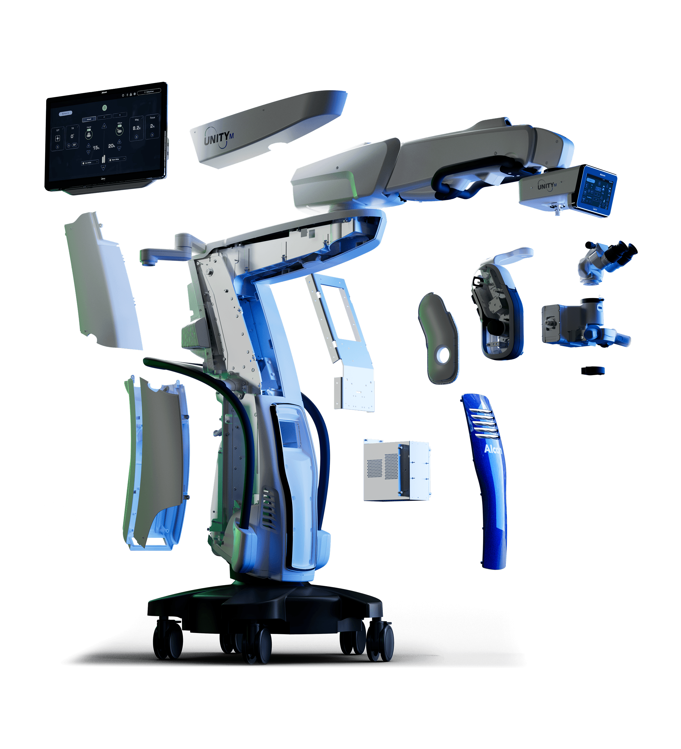
Enhanced ergonomics for ease of use in the OR4*
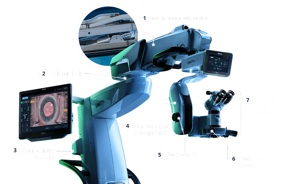
1. Electromagnetic brake—easier to position the microscope in place.18
2. Column lock—instantly locks position for one-touch stability.
3. Connectivity Module
4. Internal cable management—optimized routing with improved accessibility.4
5. Electronic tilt—adjusts the optical head to a preset or customized angle; ease of use for MIGS procedures.4
6. Redesigned handles—better ergonomics and working space.4,19*
7. Reduced stack height—supports comfort during procedures.4
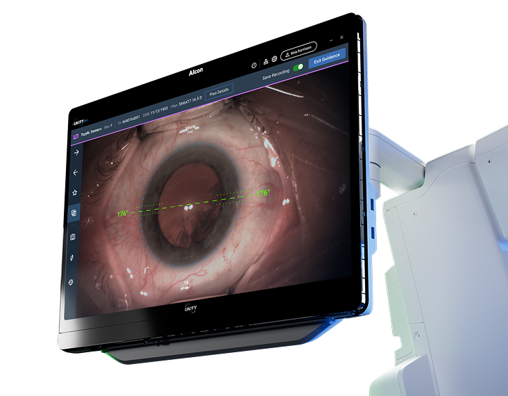
Integrate Image Guidance to support advanced visualization at every step of the surgical procedure20-23§§
Service and support
UNITY® M is supported by Alcon’s industry-leading services designed with and for your practice. Every step of the way is supported by technical and clinical services.
Technical Services
Highly responsive remote and on-site installation support, maintenance, and repair
Improve Reliability by optimal equipment setup and maintenance
Peace of Mind with industry leading service support and response times
Drive Business Predictability with proactive scheduled maintenance and fixed service costs
Clinical Services
A tailored approach to training and support empowers your practice to maximize clinical outcomes
Increase Confidence by minimizing clinical escalation and challenges, building trust in your practice
Optimize Performance by adopting advanced technology quicker & easier, providing better patient outcomes
Maximize User Proficiency through tailored training delivered by an expert clinical professional team, to help you master your equipment

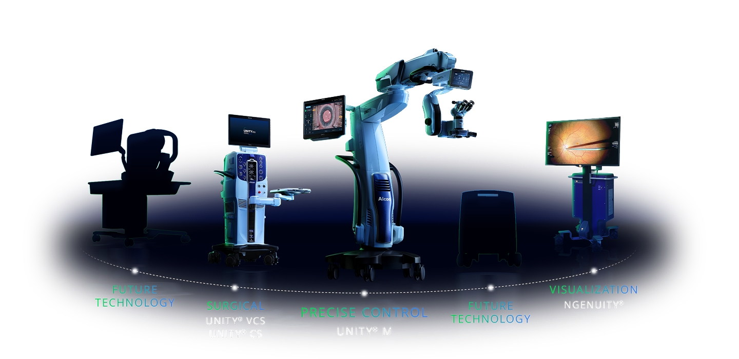
Discover the Alcon Equipment Ecosystem
Connect UNITY® M to the equipment your practice already runs
UNITY® M is the latest innovation offering precise control alongside Alcon's evolving portfolio of next-generation technologies.
UNITY® M: Frequently Asked Questions
UNITY® M is Alcon’s next-generation ophthalmic surgical microscope platform, designed to provide ophthalmic surgeons with precise control through advanced visualization technologies, customizable illumination, and a platform designed to evolve with future innovations.1,4,7,16
UNITY® M combines Color Tunable Illumination, Superior Lens Position, and advanced optical performance to deliver a stable visual experience and greater control throughout ophthalmic surgery.1,2,16‖‖
Color Tunable Illumination is an Alcon technology that is unique to UNITY® M which enables independent control of coaxial and oblique illumination, allowing surgeons to adjust the characteristics of the illumination to optimize contrast and visibility of ocular tissues. By providing greater control over how structures are illuminated, surgeons can tailor visualization to their preferences and the needs of the procedure.2,7,16††
Superior Lens Position is a proprietary Alcon optical design that positions the objective lens above the illumination module. This helps deliver a bright, stable red reflex and enhanced depth of field, supporting consistent visualization throughout surgery.1‖‖
UNITY® M combines Superior Lens Position technology with advanced illumination design to help maintain a bright, stable red reflex throughout surgery.1,16‖‖ Enhanced depth of field helps keep structures in focus across changing surgical conditions,2‖ while the expansive red reflex is designed to be less susceptible to changes in eye movement, microscope position, and pupil size.1,11-13‖ Color Tunable Illumination helps you see what matters; Superior Lens Position helps you keep it in view.
Yes. UNITY® M is designed to integrate and work with current Alcon image guidance platforms. Together, UNITY® M and integrated Image Guidance technologies create a connected workflow that helps bring diagnostic and surgical planning information into the operating room environment.
Yes. UNITY® M was developed as an upgradeable platform, allowing additional technologies and capabilities to be added as they become available.4
UNITY® M was designed to support the integration of digital visualization as part of the platform’s evolution, providing customers with flexibility as new capabilities become available.
Contact your Alcon representative to learn more about available configurations, future upgrade paths, and integration options for your surgical practice. Schedule a demo to experience UNITY® M for yourself.
Experience the Power of Precise Control1-3
Contact your Alcon representative to see UNITY® M in action.
Experience the Power of Precise Control1-3
Contact your Alcon representative to see UNITY® M in action.
IMPORTANT PRODUCT INFORMATION
Please refer to relevant product directions for use for complete list of indications, contraindications, and warnings.
*Compared to LuxOR® Revalia® microscope.
†WD = 175mm, binocular 12.5x.
‡At 50% illumination; 5.1 minutes with UNITY® M, 1.1 minutes with Zeiss Lumera§ Xenon (focused beam illumination).
§Trademarks are the property of their respective owners.
‖Vs. traditional microscopes.
¶Red reflex area: UNITY® M 2001.29 mm2 vs. focused beam 316.20 mm2.
**Compared to existing ophthalmic surgical microscopes as of May 2026.
††Based on laboratory evaluation of USAF charts and red reflex intensity versus Zeiss Lumera§.
‡‡For coaxial light.
§§Compared to analog microscopes including the Leica Proveo§ 8 and Zeiss OPMI Lumera§ 700 scopes.
‖‖Superior positioning of the objective lens relative to the light source.
References:
1. Alcon Data on File, 2026. [REF-29340].
2. Alcon Data on File, 2026. [REF-29342].
3. Alcon Data on File, 2026. [REF-29354].
4. UNITY® M User Manual.
5. Jivrajka R, Shammas MC, Boenzi T, Swearingen M, Shammas HJ. Variability of axial length, anterior chamber depth, and lens thickness in the cataractous eye. J Cataract Refract Surg. 2008;34(2):289-294.
6. ZEISS Opmi Lumera§ 700 User Manual.
7. Alcon Data on File, 2026. [REF-29343].
8. Alcon Data on File, 2026. [REF-29341].
9. Aydin B, Dinç E, Yilmaz SN, et al. Retinal endoilluminator toxicity of xenon and light-emitting diode (LED) light source: rabbit model. Cutan Ocul Toxicol. 2014:33(3);192-196.
10. Alcon Data on File, 2026. [REF-29411].
11. Cionni RJ, Pei R, Dimalanta R, Lubeck D. Evaluating Red Reflex and Surgeon Preference Between Nearly-Collimated and Focused Beam Microscope Illumination Systems. Transl Vis Sci Technol. 2015;4(4):7.
12. Alcon Data on File, 2014. [REF-00728].
13. Alcon Data on File, 2019. [REF-24564].
14. Alcon Data on File, 2026. [REF-29410].
15. Alcon Data on File, 2026. [REF-29753].
16. Alcon Data on File, 2026. [REF-29355].
17. Lubeck DM. Red reflex stability improves illumination. Ocular Surgery News US Edition. Healio.com/Ophthalmology. November 25, 2013.
18. Alcon Data on File, 2026. [REF-30087].
19. LuxOR® Revalia® Operator’s Manual.
20. Kim YJ, Kim YJ, Nam DH, et al. Contrast, visibility, and color balance between the microscope vs. intracameral illumination in cataract surgery using a 3D visualization system. Indian J Ophthalmol. 2021;69;927-931.
21. Ehlers JP, Uchida A, Srivastava SK. Combining intraoperative optical coherence tomography and 3D digital visualization for vitreoretinal surgery in the DISCOVER study. Retina. 2018;38:S88-S96.
22. Alcon Data on File, 2017. [REF-02147].
23. Franklin AJ, Sarangapani R, Yin L, Tripathi B, Riemann C. Digital vs analog surgical visualization for vitreoretinal surgery. Retinal Physician. 2017;14(May):34-36, 38-40.