

Vision Surgery
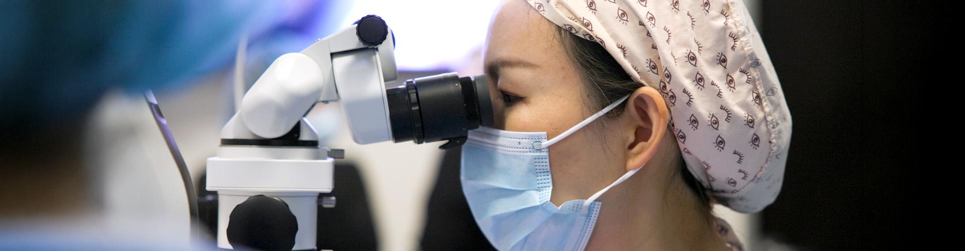
Cataract and refractive surgeons describe the procedural moments when UNITY® M, an ophthalmic microscope, first changed what they could see and how they responded in real time.
In cataract surgery, visualization is not simply about seeing the surgical field. It shapes how a surgeon moves through the case, responds to subtle changes in anatomy and makes decisions in real time. From the moment the microscope comes into focus, the quality and consistency of that view can influence what the surgeon sees, and what happens next.1
That is why advances in surgical microscopy matter. When some of the country’s leading surgeons recently performed their first procedures with the UNITY® M ophthalmic microscope, the impact was apparent the moment they looked through the oculars. A broad range of anatomy remained in focus, fine tissue details were easier to distinguish and the red reflex stayed stable as surgical conditions changed.2-5,*
These are the moments when microscope innovation moves from an engineering advancement to a surgical tool — helping support the visualization, control and decision-making that matter most throughout a procedure. Let’s hear more about what the surgeons had to say.
What Surgeons Notice First When Using UNITY® M
Across interviews, surgeons pointed to several common observations from their first cases with UNITY M:
- • Deep depth of field across multiple anatomical planes3,*
- • Enhanced contrast and detail recognition during surgery2-4
- • Less need to adjust focus during a procedure3,†
- • Greater visibility of subtle tissue details2-4
- • Stable visualization as the eye moved or surgical field changed3
- • A clear monitor image for surgical staff and observers
For Dr. Robert Melendez, a cataract and refractive surgeon at Juliette Eye Institute in New Mexico, the difference was apparent as soon as he looked through the oculars.
“What really excited me most was the visualization. It was probably the best I’ve ever seen in my career as a surgeon,” he said.
Dr. Eric Rosenberg, a cornea, cataract and refractive surgeon at SightMD in New York, also entered the operating room unsure how much ophthalmic visualization could improve.
“To be honest with you, I was skeptical going into the OR because I wasn’t sure that I was going to be that impressed,” he said. “But I was blown away with UNITY M.”
How Depth of Field Supports Surgical Control
Among those first impressions, depth of field emerged as one of the most consistent — and consequential — differences surgeons described.
Dr. William Trattler, a cornea, cataract and refractive surgeon at the Center of Excellence in Eye Care in Miami, said UNITY M’s depth of field helped him maintain focus across multiple parts of the eye.3,*
“What I found most impressive was the huge depth of field,” he said. “I was able to see and focus from the capsule all the way up to the cornea.”
For Dr. Trattler, the depth of field meant fewer focus adjustments as he moved through different phases of a procedure—particularly in an ICL case, where he described being able to visualize the depth of his entry blade, the position of the lens and haptics relative to the iris, and viscoelastic strands during removal.3
“It made it very simple to be able to focus on each surgical step without adjusting my focus point,” he said.
Dr. Dagny Zhu, a cornea and cataract surgeon practicing at Zhu Vision in Southern California, described a similar experience during her first procedure with UNITY M.
“I feel like I can see more planes at once,” she said. “It offers a greater depth of focus, so I wasn’t constantly focusing in and out.”
That experience was particularly relevant during a complex first case involving a dense cataract and high myopia. She noted that as the iris-lens diaphragm moved posteriorly during phacoemulsification, the broader range of focus helped her continue working without repeatedly searching for a new focal plane.3
“UNITY M allowed me to see that greater range without having to continue to find focus,” Dr. Zhu said. “It made the surgery a lot more straightforward.”
How Visualization Influences Surgical Decision-Making
Visualization can be especially important when a surgeon sees a small detail that changes what happens next.6 Across their first cases, surgeons described moments when clearer visualization helped them recognize subtle findings, reassess anatomy, and adjust their approach in real time.
During one case, Dr. Zhu noticed subtle Descemet’s folds near an entry site and was able to modify how she inserted her instruments into the eye.
“Catching potential problems early is huge,” she said. “It’s not about doing perfect surgery every time. It’s about catching those small things and having the ability to fix them and prevent it from being an issue down the line.”
Her experience illustrates a practical link between visualization and intraoperative decision-making: seeing a small tissue change can inform the next maneuver. Dr. Jennifer Loh, a cataract and refractive surgeon at Loh Ophthalmology Associates in Miami, similarly connected depth of field and clarity with identifying findings that could affect the course of a procedure.
“Having that depth of field can help you identify a possible posterior capsular tear or may help you see a small, retained lens fragment that otherwise, you may have missed,” she said.
For Dr. Rosenberg, adjustable color profiles offered another way to evaluate anatomy during different stages of surgery.
“We went to every color module to see what highlighted best at each step, including incisions, capsulotomy, phacoemulsification, cortex removal and posterior clean up,” he said.
Maintaining Visualization During Unexpected Movement
The need for consistent visualization became even more apparent when conditions changed unexpectedly. In several first cases, patient movement tested whether the surgical view could remain stable without interrupting the surgeon’s focus.
Dr. Trattler described one case in which a patient moved unexpectedly during surgery.
“The microscope has such great depth of field and wide access,” he said. “I was able to keep a lot of it in focus even after they moved.”
Dr. Rosenberg similarly noted that the red reflex remained stable as the patient moved.5
“Even as they were moving, we never lost a red reflex,” he said. “It was phenomenal. It made my job easier.”
For Dr. Melendez, the consistency meant he could focus less on managing the microscope and more on the procedure itself.
“It just felt like I was doing the surgery,” he said. “I wasn’t even thinking about the microscope.”
A Clearer View for the Surgical Team
The visualization experience also extended beyond the surgeon. As UNITY M supported a consistent view through the oculars, the monitor image also gave surgical staff and observers a clearer view of the procedure as it unfolded.
At Dr. Zhu’s practice, staff and patient family members followed the procedure on a large screen. During posterior capsule power washing, her long-time scrub technician reacted to seeing the fine capsule folds and debris removal on screen
“‘Wow, Dr. Zhu, I didn’t realize that’s what you were doing this whole time,’” she said recalling her technician’s reaction. “She was fascinated.”
Dr. Melendez described a similar reaction from his team.
“The monitor image was also super clear,” he said. “It’s the best I’ve seen in my more than 20 years of practice.”
Across these first-use experiences, the value of visualization became most apparent in the moments when it informed what surgeons did next — whether adjusting focus, modifying instrument entry, assessing the capsule, responding to movement or selecting an illumination profile for a specific step.
That is where microscope innovation mattered most: not as a specification, but as visualization designed to support precise control when the surgical view — and the next decision — were paramount.2,3,5
1. What is UNITY M?
UNITY M is a premium surgical microscope, delivering precise control to ophthalmic surgeons through advanced visualization technologies designed to provide stable, consistent views throughout surgery.2,3,5
2. How does UNITY M support patient safety and surgical outcomes?
UNITY M supports patient safety and surgical outcomes via a lower light intensity compared to focused-beam illumination microscopes to reduce the risk of phototoxicity, with a maximum exposure time 4.6x longer than Zeiss^ Lumera^.7,8,‡ The microscope is also designed to provide a stable red reflex with nearly collimated light that is 6x larger than conventional systems. Even when the eye moves off axis, the reflex still shines through.1,5,9,††
3. What do surgeons notice first when using UNITY M?
Surgeons most frequently highlighted deep depth of field, contrast and detail recognition, red reflex stability and the ability to maintain focus across multiple anatomical planes.2-5*
4. During which procedural moments did surgeons say visualization mattered most?
The interviewed surgeons discussed incision entry, capsulotomy, phacoemulsification, cortex removal, posterior cleanup and assessment of the capsule and other anatomy during surgery.
5. How can UNITY M support surgical control?
Surgeons described fewer focus adjustments and visibility across multiple planes.3* UNITY M is designed to support stable red reflex as eye position changes and deep depth of field as the eye shifts or the chamber changes.3,5
For these surgeons, the first case changed their expectations. What stood out was not simply the microscope itself, but how visualization supported them through key moments of the procedure.2,3,5
Jennifer Loh, MD
Dr. Jennifer Loh is a board-certified comprehensive ophthalmologist practicing in South Miami, specializing in cataract and refractive cataract surgery. Her practice also includes the management of dry eye disease, pterygium, glaucoma and comprehensive eye care. Dr. Loh performs cataract surgery using technologies including femtosecond laser and advanced intraocular lenses. She has been recognized as a Premier Surgeon Top 300 Innovator in Refractive Cataract Surgery.
Dr. Loh graduated summa cum laude from Butler University and earned her medical degree from Indiana University School of Medicine, where she also completed her ophthalmology residency. During medical school, she was inducted into the Indiana University chapter of the Gold Humanism Honor Society. She also served as chief resident during both her intern year and ophthalmology residency.
Robert F. Melendez, MD, MBA
Dr. Robert Melendez is a board-certified ophthalmologist specializing in cataract and refractive surgery and founder of Juliette Eye Institute in New Mexico. He has performed more than 15,000 cataract and refractive surgeries.
Dr. Melendez earned his bachelor’s and master’s degrees in biochemistry, medical degree and MBA from the University of New Mexico (UNM). He completed his ophthalmology residency at The University of Texas Health Science Center at San Antonio, where he served as Chief Resident and was voted resident eye surgeon of the year.
Alongside his clinical work, Dr. Melendez has held several leadership and educational roles with the American Academy of Ophthalmology (AAO). He served as Chair of AAO’s Young Ophthalmologist Committee, Editor-in-Chief of the ONE Network and Secretary for Online Education, overseeing the ONE Network and EyeWiki.org.
Dr. Melendez is also founder and president of the Juliette RP Vision Foundation, supporting visually impaired college students and retinal research. He previously served as a flight surgeon and Lieutenant Colonel in the United States Air Force/New Mexico Air National Guard.
Eric D. Rosenberg, DO
Dr. Eric D. Rosenberg is a fellowship-trained ophthalmologist specializing in cornea, cataract and refractive surgery. He practices at SightMD, serving patients across Long Island, New York.
Dr. Rosenberg provides anterior segment care, including treatment for corneal disease, cataracts and ocular surface disorders, as well as LASIK and PRK laser vision correction. He also specializes in modern cataract surgery and provides glaucoma care.
Alongside his clinical work, Dr. Rosenberg is an active researcher and educator. He has published more than 30 peer-reviewed journal articles, six book chapters and the ophthalmology textbook Operative Dictations in Ophthalmology, a resource used by residents and fellows to learn surgical techniques. His research focuses on cornea, refractive surgery and external eye diseases.
Dr. Rosenberg has been recognized among Ophthalmology Management’s “40 Under 40,” the “Rising Stars 100 Power List” and as a New York Super Doctors Rising Star for five consecutive years. He also serves as a peer reviewer for major ophthalmology journals.
William B. Trattler, MD
Dr. William B. Trattler is a board-certified ophthalmologist specializing in corneal, cataract and refractive surgery at the Center for Excellence in Eye Care in Miami. He lectures nationally on cataract and refractive surgery, corneal disease and other ophthalmic topics. Dr. Trattler earned his bachelor’s degree from Dartmouth College and medical degree from the University of Miami School of Medicine. He completed his ophthalmology residency at the University of Pennsylvania’s Scheie Eye Institute, followed by fellowship training in cornea and refractive surgery at the University of Texas Southwestern Medical Center.
Alongside his clinical work, Dr. Trattler has served as an investigator for ophthalmic technologies and treatments, including corneal collagen crosslinking for keratoconus and post-LASIK ectasia. He also serves on the volunteer faculty at Florida International University’s Herbert Wertheim College of Medicine and the University of Miami’s Bascom Palmer Eye Institute.
Dr. Trattler has authored numerous articles and abstracts and co-authored the textbook Microbiology Made Ridiculously Simple. His professional recognitions include being named among The Ophthalmologist’s “Top 100” most influential people in ophthalmology and an Ocular Surgery News Premier Surgeon 300 Innovator in Refractive Cataract Surgery.
Dagny Zhu, MD
Dr. Dagny Zhu is a board-certified ophthalmologist specializing in cornea, cataract and refractive surgery.
She is Medical Director and Founder of Zhu Vision – Modern Laser & Cataract Eye Surgery in Orange, California, and holds a clinical faculty appointment at the Western University School of Osteopathic Medicine.
Dr. Zhu leads FDA clinical research trials in cataract, refractive, presbyopia and keratoconus treatments and educates ophthalmic surgeons on surgical procedures and technologies. She has delivered or been featured in more than 300 lectures, scientific presentations, publications and press articles.
Dr. Zhu earned her medical degree from Harvard Medical School and completed her ophthalmology residency at the Doheny Eye Institute/University of Southern California and a cornea and refractive surgery fellowship at Bascom Palmer Eye Institute.
She holds leadership positions with the American Academy of Ophthalmology and American Society of Cataract and Refractive Surgery and serves on the editorial boards of ophthalmology publications. Her professional recognitions include The Ophthalmologist’s “Rising Star” Power List, Ophthalmology Management’s “Top 40 Under 40” and Castle Connolly “Top Doctor.”
*Compared to traditional microscope
†DoF at 7x: UNITY M 3198.1 µm vs. Zeiss^ Lumera 2813.0 µm & 18x: UNITY M 1336.8 µm vs. Zeiss^ Lumera 946.9 µm. WD=175mm, binocular 12.5x.
‡At 50 % illumination; 5.1 minutes with Unity M, 1.1 minutes with Zeiss Lumera† Xenon (focused beam illumination).
††Red reflex area: UNITY M 2001.29 mm2 vs. focused beam 316.20mm2
^Trademarks are the property of their respective owners.
1. Cionni RJ, Pei R, Dimalanta R, Lubeck D. Evaluating red reflex and surgeon preference between nearly-collimated and focused beam microscope illumination systems. Transl Vis Sci Technol. 2015;4(4):7. doi:10.1167/tvst.4.4.7
2. Alcon Data on File, 2026. REF-29354.
3. Alcon Data on File, 2026. REF-29342.
4. Alcon Data on File, 2026. REF-29355.
5. Alcon Data on File, 2026. REF-29340.
6. Han SB, Liu YC, Mohamed-Noriega K, Mehta JS. Application of Intraoperative Optical Coherence Tomography Technology in Anterior Segment Surgery. J Ophthalmol. 2022;2022:1568406. Published 2022 Apr 8. doi:10.1155/2022/1568406.
7. ZEISS Opmi Lumera 700 User Manual
8. Alcon Data on File, 2026. REF-29343.
9. Alcon Data on File, 2026. REF-29411.
© 2026 Alcon Inc. US-UNM-2600043