


THIS IS STABILITY.
The HYPERVIT® blade vitrectomy probe.*
Optimize performance with 20,000 cuts per minute.1-3


Superior Fluidic Stability1-3*
Combined with valved cannulas and CONSTELLATION® Vision System’s IOP compensation, the HYPERVIT® Dual Blade Bevel vitrectomy probe enables stable, closed-system intraocular surgery4,5
*HYPERVIT® Dual Blade Vitrectomy Probe compared at 20,000 CPM (maximum cut rate in core mode) with Advanced ULTRAVIT® Probe at 10,000 CPM (maximum cut rate in core mode)
Reduced Fluidic Turbulence
ULTRAVIT® probe
Advanced ULTRAVIT® probe
closer port proximity to retina with improved efficiency
HYPERVIT® Dual Blade probe
closer port proximity to retina with maximum efficiency & tissue stability

HYPERVIT® features a continuously open port that reduces fluidic turbulence.
Reduced Traction
Dual Blade design with 20,000 CPM enables reduced pulsatile traction during procedures.1,2
Higher vitreous flow rate with HYPERVIT® Probe1


†HYPERVIT® Dual Blade Vitrectomy Probe compared at 20 000 CPM (maximum cut rate in core mode) with Advanced ULTRAVIT® Probe at 10000 CPM (maximum cut rate in core mode); 95% confidence interval, n= 6 to 12 probes (27G), n=8 (25G)
Reduced Backflow
The dual-cutting 20,000 CPM HYPERVIT® probes showed less back flow (retropulsion) and improved intraoperative flow stability.3

HYPERVIT® is designed with a beveled shape to decrease the size of the dead recirculatory flow zone inside the tip, allowing for reduced backflow.
Enhanced Vitreous Removal Efficiency
Improved Vitreous Flow
The continuously open port in the 20,000 CPM dual-pneumatic drive helps to improve vitreous flow rate.6,7*

*HYPERVIT® Dual Blade Vitrectomy Probe compared at 20,000 CPM (maximum cut rate in core mode) with Advanced ULTRAVIT® Probe at 10,000 CPM (maximum cut rate in core mode)
Vitreous Removal Efficiency
At 20,000 CPM, HYPERVIT® has been shown to offer a higher, more efficient vitreous flow rate.6,7
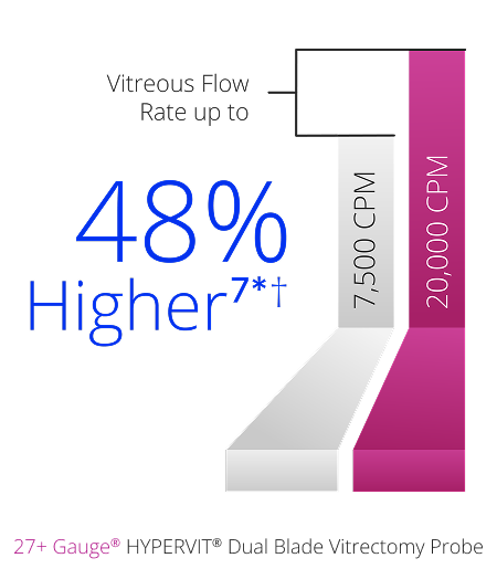
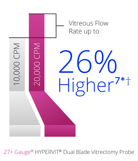
*HYPERVIT® Dual Blade Vitrectomy Probe 27+® Ga compared at 20 000 CPM (maximum cut rate in core mode) with Advanced ULTRAVIT® Probe 27+® Ga at 10 000 CPM (maximum cut rate in core mode) and ULTRAVIT® Vitrectomy Probe 27+® Ga at 7500 CPM (maximum cut rate in core mode)
†95% confidence interval, n = 6 to 12 probes
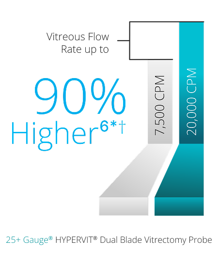
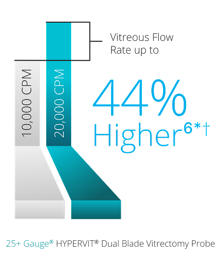
*HYPERVIT® Dual Blade Vitrectomy Probe 25+® Ga compared at 20 000 CPM (maximum cut rate in core mode) with Advanced ULTRAVIT® Probe 25+® Ga at 10 000 CPM (maximum cut rate in core mode) and ULTRAVIT® Vitrectomy Probe 25+® Ga at 7500 CPM (maximum cut rate in core mode)
†95% confidence interval, n = 8 probes
Dual Pneumatic Drive
The dual pneumatic drive feature enables efficient aspiration and precise shearing at any cut rate.8

Improved Surgical Versatility
Closer proximity to the retina
The bevel tip design of HYPERVIT® allows for improved access to tissue plane due to reduced port-to-surface distance.9*
*HYPERVIT® Dual Blade Vitrectomy Probe compared with ULTRAVIT® Probe
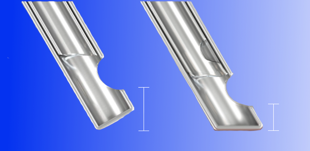


*HYPERVIT® Dual Blade Vitrectomy Probe compared with ULTRAVIT® Probe
Clinical Support
Instructions for Use (IFU)
For a full list of indications, contraindications and warnings, please visit ifu.alcon.com and refer to the relevant product’s instructions for use.
Alcon Experience Academy
For relevant training content from industry thought leaders
*Ultravit / Hypervit Vitrectomy Probe Pak with Engauge RFID. Directions for use.
References:
1. Alcon Data on File, 2018. [REF-10328].
2. Irannejad A, Tambat S, Abulon DJK. Retropulsion and mass flow of 27-gauge vitrectomy probes comparison of dual-blade flat-tipped probes and single blade beveled probes. Poster presented at 18th Congress of the European Society of Retina Specialists; September 20–23, 2018; Vienna, Austria.
3. Novack R, Zhou J, Abulon DJK, Buboltz DC. Relationship of duty cycle versus cut rate for two commercially available vitrectomy systems. Poster presented at 28th Annual Meeting of the American Society of Retina Specialists; August 28–September 1, 2010; Vancouver, BC, Canada.
4. Abulon DJ, Charles M, Charles DE. Globe stability during simulated vitrectomy with valved and non-valved trocar cannulas. Clin Ophthalmol. 2015;91745–1752.
5. Alcon Data on File, 2018. [REF-00405].
6. Alcon Data on File, 2018. [REF-01617].
7. Riemann CD, Zhou J, Buboltz DC. Vitreous cutter velocities dual pneumatic drive vs. single pneumatic drive with spring return probes. Poster presented at 2011 Annual Meeting of the Association for Research in Vision and Ophthalmology; May5, 2011; Fort Lauderdale, FL.
8. Alcon Data on File, 2017. [REF-10853].
Please refer to the relevant product direction for use for list of indications, contraindications and warnings.